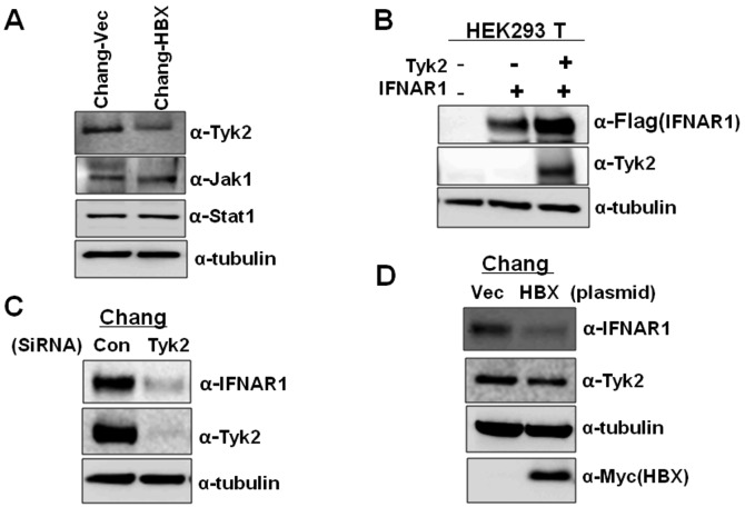
Figure 3.
HBX-mediated decrease of Tyk2 causes a reduction in IFNAR1 levels. (A) Cell lysates from Chang-Vec and Chang-HBX cells were electrophoretically separated on 10% SDS-PAGE. Tyk2, Jak1 and Stat1 were detected with the corresponding antibodies. (B) HEK293 T cells were transfected with a Flag-tagged IFNAR1 vector (1 μg) with or without a Tyk2 expression vector (1 μg) using the calcium phosphate precipitation method. Two days post-transfection, cells were harvested and the cell lysates separated on 10% SDS-PAGE followed by immunoblotting with anti-Flag and anti-Tyk2 antibodies. (C) Chang cells were transfected with Tyk2 siRNA (60 nM) and control siRNA (60 nM). Two days post-transfection, cells were harvested and the cell lysates were separated on 10% SDS-PAGE followed by immunoblotting with anti-IFNAR1 and anti-Tyk2 antibodies. (D) Chang cells were transfected with a Myc-tagged HBX vector (2 μg) and endogenous IFNAR1 and Tyk2 were detected by immunoblotting 2 days post-transfection.

