
Figure 4.
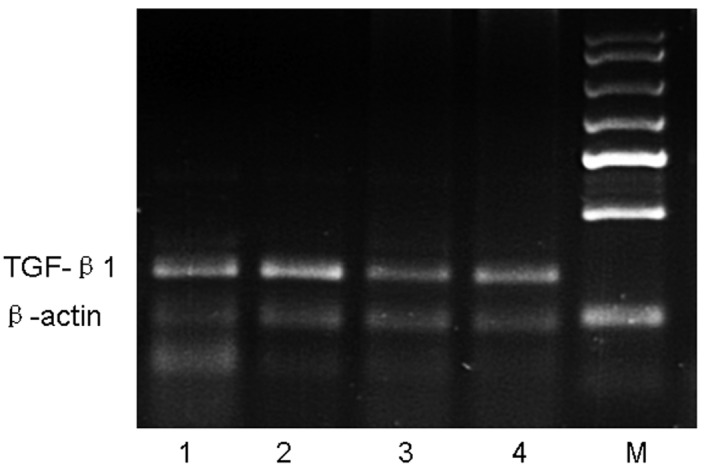
RT-PCR analysis of TGF-β1 mRNA in lung tissue. Mice were sacrificed 24 h after the final OVA challenge, and mRNA was then isolated and subjected to semi-quantitative RT-PCR analysis of TGF-β1. Expression of β-actin was used as a loading control. Lane M, marker; lane 1, control group; lane 2, asthmatic group; lane 3, astragalus extract-treated group; lane 4, budesonide-treated group.