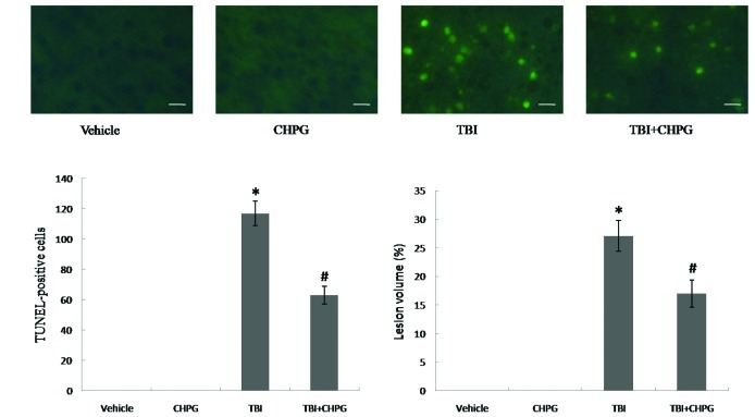
Figure 2.
Administration of CHPG attenuated neuronal damage after TBI in vivo. CHPG (250 nM) was injected into right lateral ventricle in a volume of 5 μl 30 min before TBI. TUNEL staining was used to detect apoptotic cells in the cortex 24 h after TBI. Sections were examined by fluorescence microscopy (A) and the number of TUNEL-positive (apoptotic) cells was counted in 5 fields in each section (B). Lesion volume was measured by cresyl violet staining 7 days after TBI (C). Scale bars, 20 μm. The data are presented as means ± SD from seven experiments. *P<0.05 vs. vehicle group; #P<0.05 vs. TBI group.

