
Fig. 5.
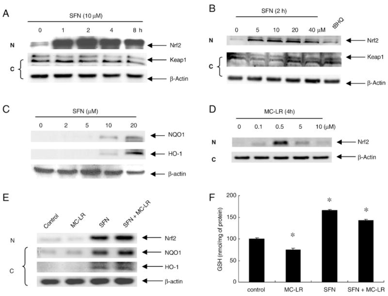
SFN effects on activation of Nrf2 pathway in HepG2 cells. (A) and (B), Nrf2 is enriched in nucleus after SFN treatment. (A) Time-course study. Nuclear (N) and cytoplasmic fractions (C) were extracted from cell lysate of HepG2 treated with 10 μM of SFN. (B) Dose-response study. The cells were treated with 0, 5, 10, 20, 40 μM of SFN for 2 h. tBHQ, a known Nrf2 pathway inducer, was used as a positive control. (C) SFN induces NQO1 and HO-1 expression. Cytoplasmic fractions were probed for NQO1 and HO-1. (D) Increase in nuclear Nrf2 protein in response to MC-LR. HepG2 cells were treated with MC-LR (0, 0.1, 0.5, 5, 10 μM) for 4 h. Cytosolic and nuclear fractions were prepared from each cell line and subjected to western blot analysis with anti-Nrf2 and β-actin antibody respectively. (E) HepG2 cells treated with 10 μM of MC-LR, 10 μM of SFN, and (SFN+MC-LR) for 4 h. Nuclear and cytoplasmic fractions were extracted from cell lysate. Nuclear fractions were probed for Nrf2, and cytoplasmic fractions were probed for HO-1 and NQO1. (F) Intracellular GSH was enhanced by SFN alone and (SFN + MC-LR) treatment. Cells treated with MC-LR alone, SFN alone and (SFN + MC-LR) for 12 h. Each value represents the mean±S.D. of three determinations. *, p<0.05.