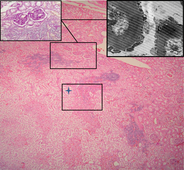
Figure 2.
Histological, histochemical and ultrastructural findings. Kidney. Cortico- medular area of one reniculum. Multifocal lymphoplamacytic interstitial nephritis. H&E (100X, bar=200 μm). Up right side inset: Higher magnification of renal cortical area showing aggregate of lymphoplasmocytic cells and two glomeruli with (PAS positive). PAS technique (400x, barr= 100 μm). Up right side inset: EM picture showing thickening of glomerular basement membranes. (Bar 2 μm, EM technique).

