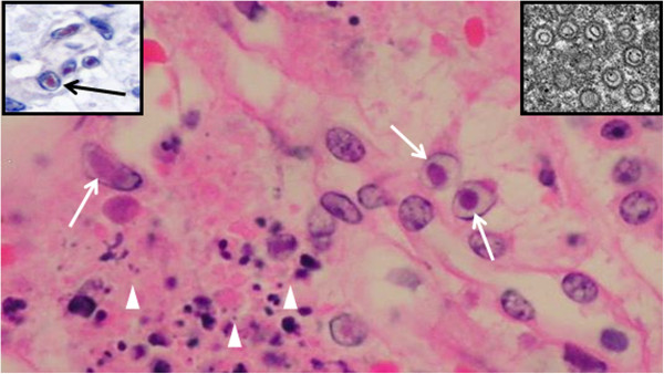
Figure 3.

Immunohistological and ultrastructural interstitial kidney findings. Kidney. Higher magnification of Figure 2 (box with star). Interstitial cell necrosis (arrowheads) and intranuclear inclusion bodies (arrows) in tubuloepithelial cells. H&E (bar=10 μm). Inset up right: Herpes virus particles (EM) (bar=10 nm). Inset up left: Inclusion bodies labeled immunpositivelly for herpesvirus antigen (red colour) (arrow). IHC counterstaining with hematoxilin (bar=10 μm).
