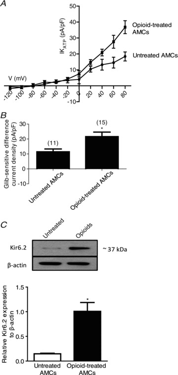
Figure 3. Upregulation of functional KATP channels and Kir6.2 subunit in opioid-treated neonatal rat AMCs.

The glibenclamide-sensitive difference current density (IKATP (pA/pF)) in untreated versus opioid-treated AMCs is plotted against voltage in the I–V plot (A), and during steps to +30 mV (B). Note the significant increase in KATP current density in opioid-treated relative to control untreated AMCs (P < 0.05 in B). C, Western blot analysis of KATP channel subunit, Kir6.2, in untreated AMCs and in AMCs cultured with combined μ-, δ- and κ-opioid agonists (2 μm) for 7 days. Note increased expression of Kir6.2 during chronic opioid exposure; β-actin was used as an internal control. Values are presented as mean ± SEM of three independent experiments (*P < 0.05).
