
Figure 3. Comparison of current–voltage relationships of a smooth muscle cell (SMC) and a PDGFRα+ cell.
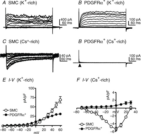
SMCs and PDGFRα+ cells expressed voltage-dependent outward currents (A and B). Depolarization evoked voltage-dependent Ca2+ currents in SMCs (C), but no inward current conductances were apparent under the same conditions in PDGFRα+ cells (D, see arrow). E and F demonstrate the summarized data of outward currents (E) and inward currents (F) from both types of cells.