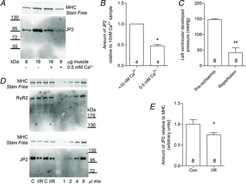
Figure 5. Junctophilin-2 is proteolysed in cardiac muscle with ischaemia–reperfusion.

A, Western blot of JP2 in rat cardiac ventricular muscle homogenized in the presence or absence of 0.5 mm free Ca2+ for 60 min at room temperature (4–15% Stain Free gel). B, mean (+SEM) amount of JP2 remaining in ventricular samples following treatments. Data are from four independent gels (*P < 0.01, Student's one-tailed paired t test). C, left ventricular developed pressure in rat hearts before and after ischaemia–reperfusion (I/R; **P < 0.001, Student's one-tailed paired t test). D, Western blots of RyR2 and JP2 in separate portions of the same homogenates of ventricular muscle from control (C) and I/R hearts (4–15 and 10% Stain Free gels). E, mean (+SEM) amount of JP2 in control (Con) and I/R hearts (*P < 0.05, Student's one-tailed unpaired t test).
