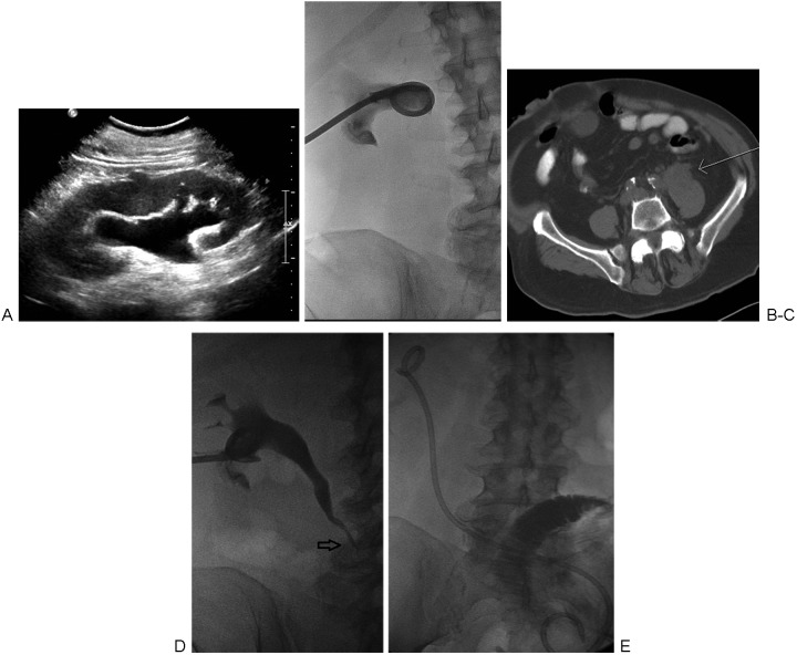
Figure 1.
(A) Ultrasound image demonstrating mild to moderate hydronephrosis of the left kidney. (B) Fluoroscopic spot image obtained following percutaneous placement of a 12F nephrostomy tube placement. Postplacement gentle injection of contrast was performed to confirm appropriate placement. (C) Axial computed tomography without contrast performed after percutaneous nephrostomy demonstrates a 4-cm retroperitoneal mass (arrow) causing the ureteral obstruction. (D) Antegrade nephrostogram performed 13 days later demonstrates a tapered narrowing of the midureter (arrow), corresponding to the location of the retroperitoneal mass. (E) The malignant stricture was traversed, and an internal ureteral stent was placed.

