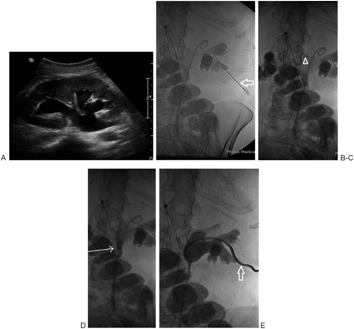
Figure 2.
(A) Ultrasound image demonstrating moderate pelvocaliectasis of the right kidney. (B) A 21-gauge needle access into posterior mid-pole calyx via a subcostal approach (arrow). Indwelling nonfunctioning ureteral stent is also noted. (C) An 0.018-inch guidewire has been advanced distally in the ureter (arrowhead). (D) Antegrade pyelogram via 6F dilator/sheath with radio-opaque tip (arrow) advanced into the proximal ureter. (E) Final image obtained following 8F percutaneous nephrostomy catheter placement (arrow).

