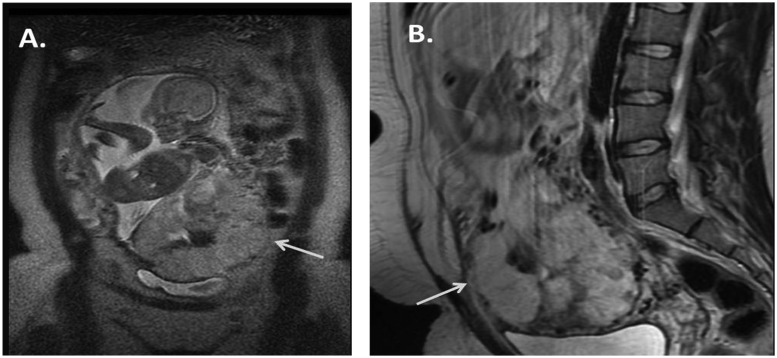
Figure 1.
(A) Coronal magnetic resonance imaging (MRI) demonstrates segment of placenta extending outside of the visible myometrium in the region of the anterior inferior abdominal wall (arrow). (B) Sagittal MRI demonstrates the placenta severely thinning the myometrium with possible extension outside of it inferiorly in the region of the bladder (arrow).

