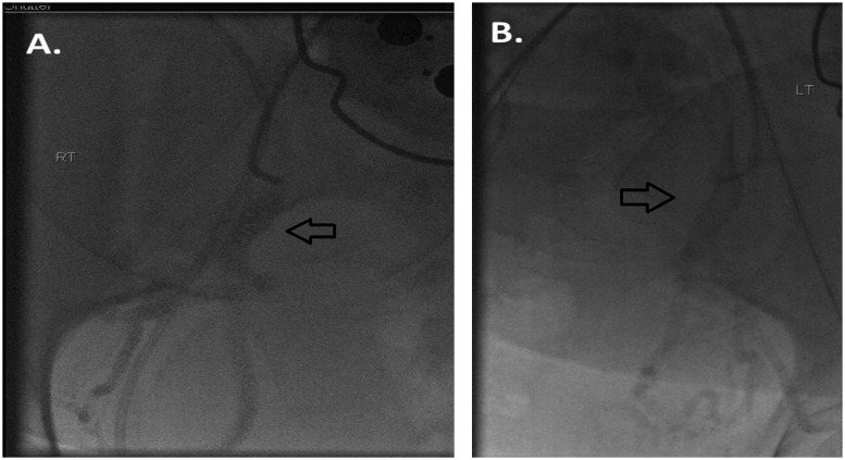
Figure 3.
(A) Following crossover access, a small test puff injection was given to document position within the right internal iliac artery. Contrast is seen within the anterior division of the internal iliac artery (arrow). (B) Following crossover access, a small test puff of contrast confirms positioning within the anterior division of the left internal iliac artery (arrow).

