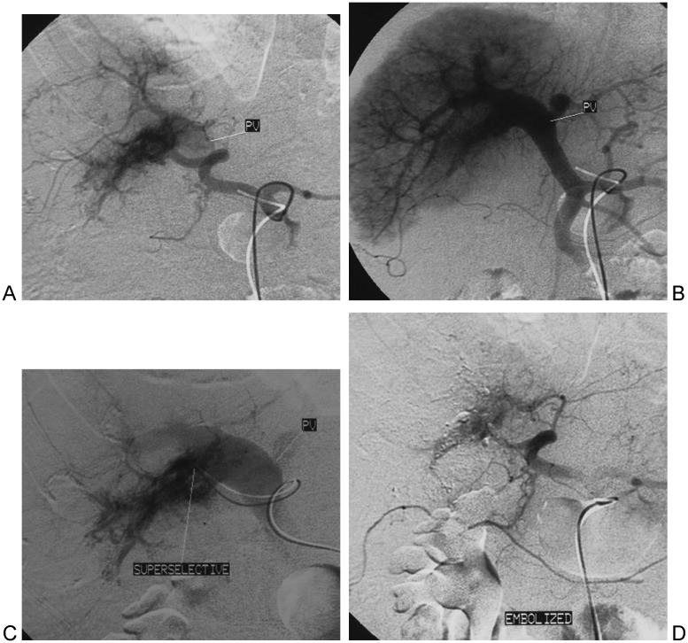
Figure 1.
High-flow arterioportal venous shunts (APVS) associated with hepatocellular carcinoma. (A) The proper hepatic artery angiogram demonstrates opacification of the main portal vein (PV) during the early arterial phase. (B) The entire portal vein (PV) is patent. (C) Preembolization superselective angiography of feeding artery to shunts. (D) Following embolization with 750 to 1000 μm polyvinyl alcohol particles, common hepatic artery angiography demonstrates complete obliteration of APVS.

