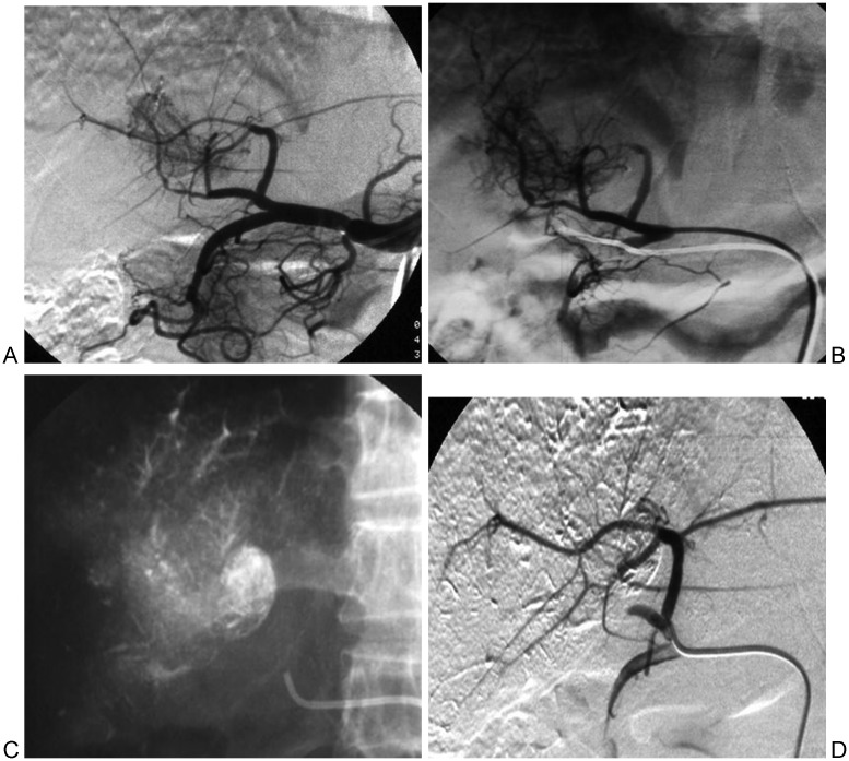
Figure 3.
Hepatocellular carcinoma with arteriohepatic venous shunts (APVS) complicated by main portal vein tumor thrombus. (A) Proper hepatic angiography demonstrates early opacification of the portal vein with thread and streak signs. (B) Superselective angiography of the middle hepatic artery. (C) Spot fluoroscopic embolization with chemotherapy lipiodol suspension and 350 to 510 μm polyvinyl alcohol particles. (D) APVS were completely occluded.

