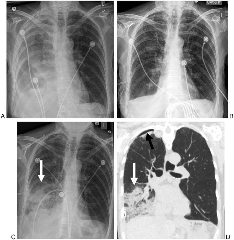
Figure 2.
An 88-year-old woman with a history of a recent myocardial infarction. (A) Frontal radiograph of the chest demonstrates a right pleural effusion. (B) Frontal radiograph of the chest immediately after chest drainage shows resolution of the right pleural effusion. (C) Frontal radiograph of the chest obtained 5 hours after chest drainage and obtained for new shortness of breath demonstrates new right lower lobe airspace opacities (arrow). (D) Coronal computed tomography scan of the chest demonstrates right lower lobe airspace disease (white arrow) consistent with reexpansion pulmonary edema. There is also a small right apical pneumothorax (black arrow).

