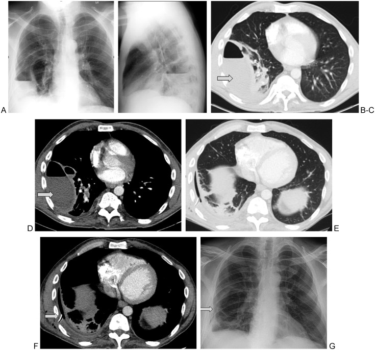
Figure 1.
45-year-old man with a history of intravenous drug abuse presented with right lower lobe pneumonia associated with a right-sided empyema. PA and lateral chest radiographs (A,B) and axial contrast enhanced computed tomography sections in lung (C) and soft tissue (D) windows reveal two septated loculated fluid collections containing air fluid levels (arrows). A 16F chest tube was placed into the larger collection under ultrasound guidance. The pleural fluid grew Streptococcus anginosis. Given the presence of thick septations, tissue plasminogen activator was administered via the chest tube with good results (E–G, arrows).

