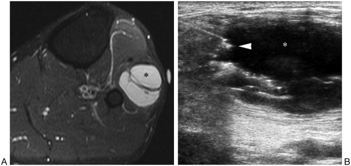
Figure 14.
Ganglion near proximal tibiofibular joint associated with peroneal nerve palsy and foot drop. (A) Fat-suppressed T2-weighted transverse magnetic resonance image of leg shows ganglion (asterisk) lateral to fibular neck. (B) Sonogram image shows needle tip (arrowhead) entering ganglion (asterisk). Subsequent aspiration of ganglion contents and steroid injection provided relief to patient.

