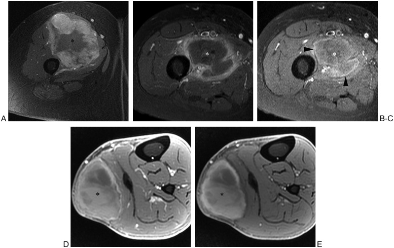
Figure 18.
Magnetic resonance (MR) images of soft tissue sarcoma, abscess, and hematoma in three different patients. (A) Fat-suppressed T1-weighted transverse MR image of thigh following intravenous administration of gadolinium chelate shows sarcoma (asterisk) in quadriceps musculature. Note the nodular peripheral enhancement, which should raise suspicion for tumor. (B) In another patient, fat-suppressed T1-weighted transverse MR image of thigh following intravenous administration of gadolinium chelate shows intramuscular abscess (asterisk) with peripheral enhancement of relatively uniform thickness. (C) Precontrast fat-suppressed T1-weighted MR image of same patient as in (B) shows high signal intensity “penumbra sign” (arrowheads) surrounding low signal intensity purulent fluid (asterisk), which has reportedly high specificity for musculoskeletal infection. (D) In another patient, fat-suppressed T1-weighted axial MR image of calf following intravenous administration of gadolinium chelate shows heterogeneous signal intensity of hematoma (asterisk) including high signal intensity components. (E) Precontrast fat-suppressed T1-weighted MR image of same patient as in (D) shows similar high signal intensity components, compatible with subacute blood products of hematoma. Follow-up MRI obtained 10 weeks later showed decrease in size of hematoma, arguing against underlying tumor.

