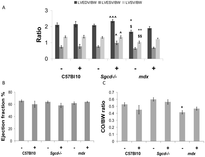
Figure 2. Effect of metoprolol treatment on left ventricular function.
(A) Left ventricular volume to body weight (BW) ratios; (B) Ejection fraction; (C) Cardiac output (CO) to body weight ratio. Reductions in left ventricular end-diastolic and (LV EDV) and left ventricular stroke volume index (LV SV) in untreated mdx mice were no longer evident after metoprolol treatment. In untreated Sgcd-/- mice there was normal left ventricular function, and after treatment with metropolol there were increases in end-diastolic, end-systolic (LV ESV) and stroke volume indexes relative to untreated mdx mice. (*different from C57 Bl10 control, $different from Sgcd-/- control, ?different from mdx control; number of symbols denotes level of significance e.g. *p<0.05, **p<0.01, ***p<0.001); (– sign indicates without metoprolol and + indicates with).

