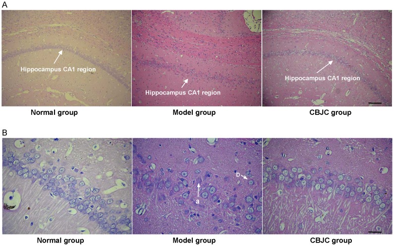
Figure 4. The histological morphology of the hippocampal CA1 region examined with HE staining.
A: photomicrographs under 100× magnification, scale bar = 40 µm; B: photomicrographs under 400× magnification, scale bar = 10 µm. In the IBO-model group, neuron arrangement is disrupted, severe lesions such as karyolysis (a) and eosinophilia (b) are observed in the nucleus and cytoplasm, and neuronal cell loss is noted.

