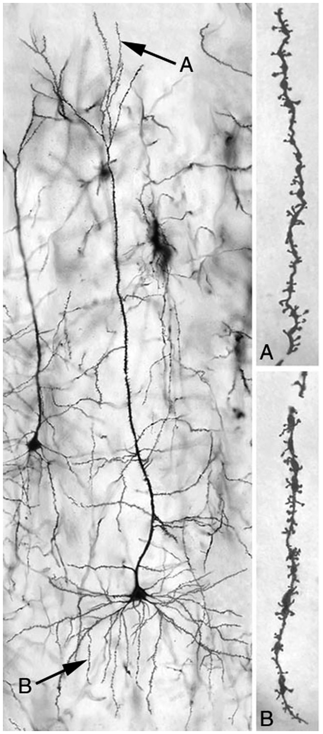
Figure 5. Photomicrograph of a typical layer 3 pyramidal cell in MPC.

“A” and “B” indicate, respectively, distal apical dendrites and third order basal dendrites, the regions from which dendritic spine density measures were obtained.

“A” and “B” indicate, respectively, distal apical dendrites and third order basal dendrites, the regions from which dendritic spine density measures were obtained.