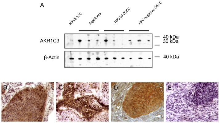
Figure 3. Analysis of AKR1C3 expression.

(A) Western blot analysis of total protein extracts from dissected tissue samples as indicated. β-Actin was used as an internal loading control (lower panel). Note the faint band of the HPV6 positive SSC while all other protein samples except for one HPV16-positive OSCC showed moderate-strong AKR1C3 expression. (B–E) Immunohistochemistry for AKR1C3 expression showing (B) strong immunostaining in a control HPV11-positive papilloma, (C) strong immunostaining in a control HPV16-positive OSCC, (D) strong immunostaining in the papilloma from 1989, (E) and no staining in the primary carcinoma from 2008. Magnification ×400.
