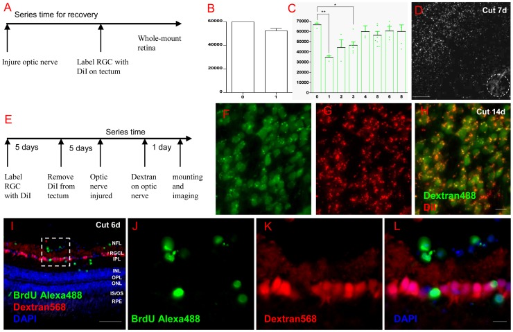
Figure 2. RGCs regenerated quickly and no new RGCs were found.
(A) Scheme of DiI labeling in RGC regeneration experiment. (B) In ONC model, over 90% of RGCs regenerated to their targets in the first week. (C) In ONT model, over 50% of RGCs regrew axons to tectum at 1 week post lesion (p<0.01, compared to normal) and the number was still significant compared to the normal animals after 3 weeks (n = 6, p<0.05). After 4 weeks, RGCs number was about 90% of normal fish and was no significant difference compared with normal (n = 3, p>0.05). (D) Periphery RGCs regrew axons to tectum more quickly than central ones during at the first week. (E) A scheme of DiI labeling RGC survival and Dextran labeling RGC regeneration in the same retina. (F-H) After 2 weeks post ONT, Dextran488 only marked RGCs were rarely found in the whole retina. (I) 6 days after ONT, BrdU+ cells could be found in the GCL and INL, but those cells did not co-localize with Dextran568 labeled RGCs. (J-L) Details of the white square in (I). Abbreviations: Neurofiber layer (NFL), retinal ganglion cell layer (RGCL), inner plexiform layer (IPL), inner nuclear layer (INL), outer plexiform layer (OPL), outer nuclear layer (ONL), photoreceptor inner segment/outer segment (IS/OS), and retinal pigment epithelium (RPE). * indicates p<0.05, ** is p<0.01. Scale bar: 100 µm (D); 50 µm (I); 10 µm (F-H, J-L).

