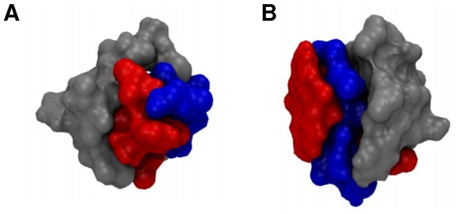
Figure 2. ApoC-II(60-70) binding preference sites on cyc(60-70).

Surface representations showing the binding preference sites for structures: (A) c1 and (B) c4. Cyc(60–70) hydrophobic face is shown in red and the hydrophilic face in blue, while the apoC-II(60–70) peptide is coloured grey.
