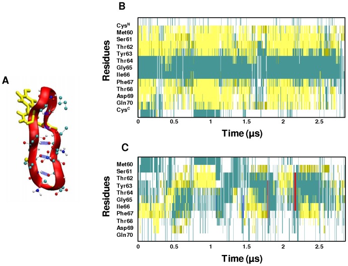
Figure 4. Structural evolution of cyc(60–70) and apoC-II(60–70).
(A) Ribbon and CPK representation of cyc(60–70) showing the persistent intra-molecular backbone hydrogen bonds in blue. The secondary structure evolution of (B) cyc(60–70) and (C) apoC-II(60–70) over 2.8 µs of simulation. The secondary structure colour codes: cyan – turn; yellow – extended conformation; green – hydrogen bridge; white – coil; blue – 3-10 helix; red – π-helix.

