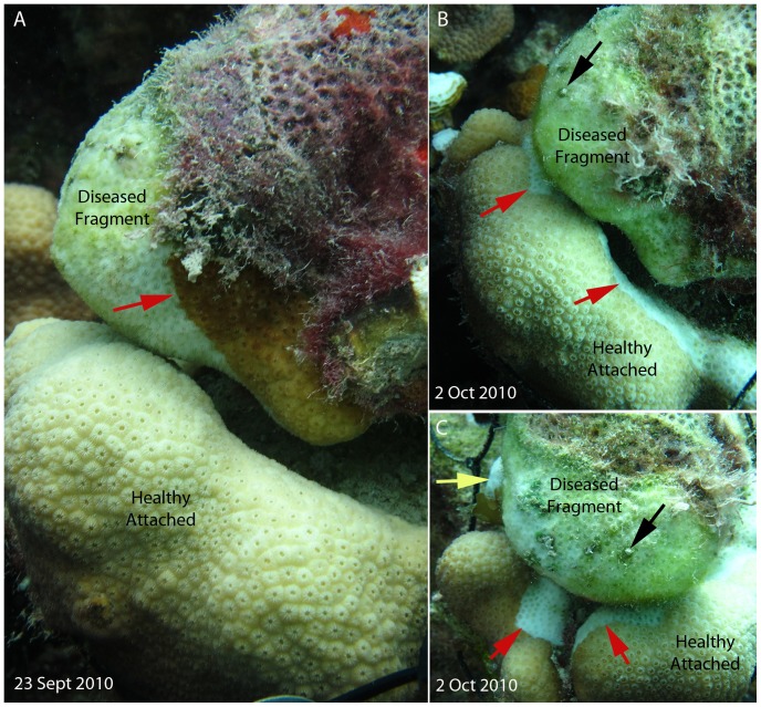
Figure 2. Example of disease transmission.
A) Experimental set up of a diseased fragment attached to an unaffected colony. Red arrow indicates active advancing lesion. B) The same experimental setup three weeks later; the diseased fragment has experienced total mortality. Red arrows indicate where a new lesion has initiated on a previously unaffected colony. C) Second view of new lesions on the previously unaffected colony. Black arrows indicate identical spot in B, red arrows as in B, and the yellow arrow indicates a small colony of Agaricia agaricites that also was recently denuded of living tissue (See Supporting Information Figure S1).

