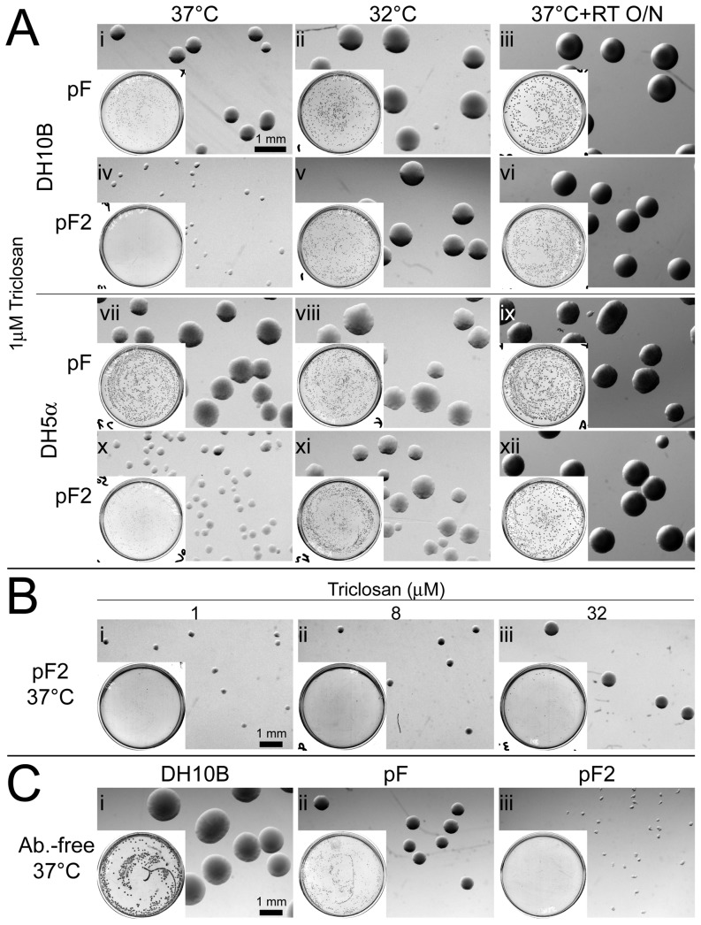
Figure 2. Impact of mfabI plasmid on host cell growth on solid support media.
(A) Growth suppression of host cells by mfabI plasmid at standard growth temperature. Shown are colonies of DH10B (i–vi) or DH5α (vii–xii) cells grown on 1 µM triclosan plates; insert is the low-magnification view of the whole plates. Cells were transformed by pF (i–iii & vii–ix) or pF2 (iv–vi & x–xii) and grown in 37°C (i, iv, vii & x) or 32°C (ii, v, viii & xi) for 24 hrs. Plates incubated in 37°C were imaged again after additional 24-hr. incubation in room temperature (iii, vi, ix & xii). (B) Rescue of mfabI plasmid-induced growth suppression by higher concentration of triclosan. pF2-transformed DH10B cells were grown in different concentration of triclosan. Equal amounts of transformed cells were plated with 1 (i), 8 (ii), or 32 µM (iii) triclosan, and incubated in 37°C for 24 hrs. (C) Triclosan resistance-independent effect of mfabI on host cell growth. Clonal untransformed (i), pF- (ii), or pF2-transformed (iii) DH10B cells were plated on antibiotic-free plates and incubated in 37°C for 24 hrs. Scale bar is 1 mm.

