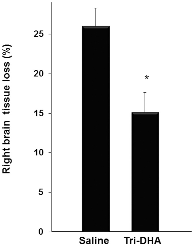
Figure 6. Long-term effect of Tri-DHA on cerebral tissue death at 8 wk after H/I.

Mice were subjected to 15-min H/I and received 2 i.p. administrations of 0.375 g Tri-DHA/kg (n = 6) vs. saline (n = 5). At 8 wk after H/I mice were sacrificed and brains were fixed with 4% paraformaldehyde and 10 µm-thick slices were cut and preserved. Nissl staining was used for identifying neuronal and brain structure. As described in Methods right brain tissue loss in relation to the contralateral hemisphere was calculated and expressed as a percentage. Each bar represents the mean ± SEM. * p<0.05.
