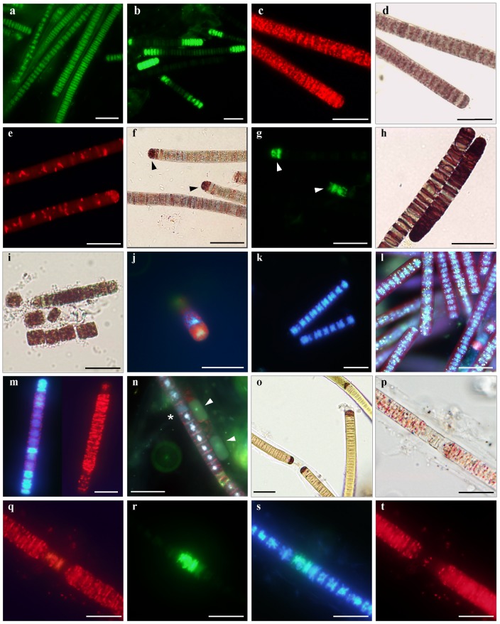
Figure 1. In situ detection of cellular functions in cyanobacterium Phormidium .
autumnale . a–l. Cultures of Phormidium autumnale 845 CCALA grown on BG-11 medium. a. SYTOX Green staining of formaldehyde-pretreated filaments; b. variable staining of live samples after 30 min incubation with 5 µM SYTOX Green; c–d. fluorescence and bright field image of CTC-stained filaments in active phase of growth; e. partially de-activated cells stained with CTC; f–g. INT-treated samples post-stained with SYTOX Green, cells that accumulated INT-formazan (f, arrowheads) were also SYTOX-positive (g, arrowheads); h. INT-stained filaments in logarithmic phase of growth; i. disintegration of filaments after 24-h incubation with CTC; j. pattern of CTC-formazan deposition; k. DAPI-stained nucleoids in living filaments; l. yellow-green metachromatic inclusions in DAPI-stained filaments viewed with a long pass emission filter; m. reduced fluorescence intensity of DAPI-stained nucleoids in cells accumulated CTC-formazan (natural samples); n. cells from old laboratory cultures simultaneously stained with DAPI, CTC and SYTOX Green under UV-illumination, extensively damaged cells lack SYTOX Green and DAPI staining of nucleoids (arrowheads) or have whole-cell DAPI signal (asterisk); o–t. Field-collected samples of mats in July and September; o. accumulation of INT-formazan by the terminal cells of filaments; p. a filament with thick sheath stained simultaneously with CTC (q), SYTOX Green (r), and DAPI (s), and showing pigment autofluorescence (t). Scale bars are 20 µm.

