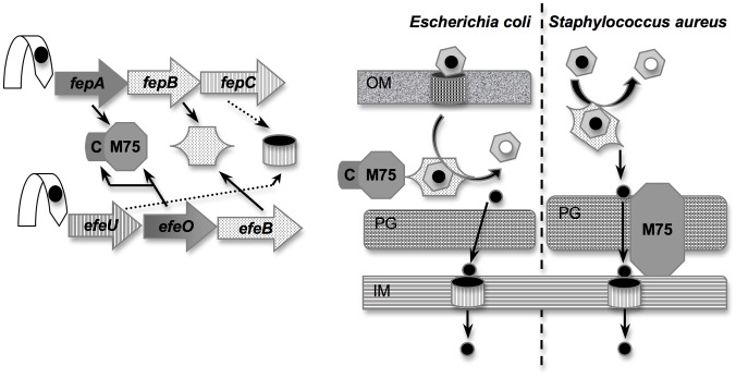
Figure 1. General organization of the EfeUOB and FepABC systems.
This scheme represents both the genetic (left part of the figure) and envelope organization (right part of the figure) of the S. aureus fepABC and E. coli efeUOB. Orthologous proteins are shown with the same pictogram and the same color. Both operons are regulated by iron loaded Fur repressor. Fur repressor is represented by an arrow with a black circle for iron; FepB and EfeB by a hexagone; FepC and EfeU by a hollow barrel; EfeO by a N-terminal square corresponding to the cupredoxin domain (C) and a C-terminal hexagone corresponding to the M75 peptidase domain; FepA which has only a M75 peptidase domain by a hexagone; heme is represented by a ring and a black circle for iron; HasR, the outer membrane receptor for heme is represented by a barrel; OM for outer membrane; PG for peptidoglycan; IM for inner membrane. Their putative function and cellular localization are shown in the right part of the figure.

