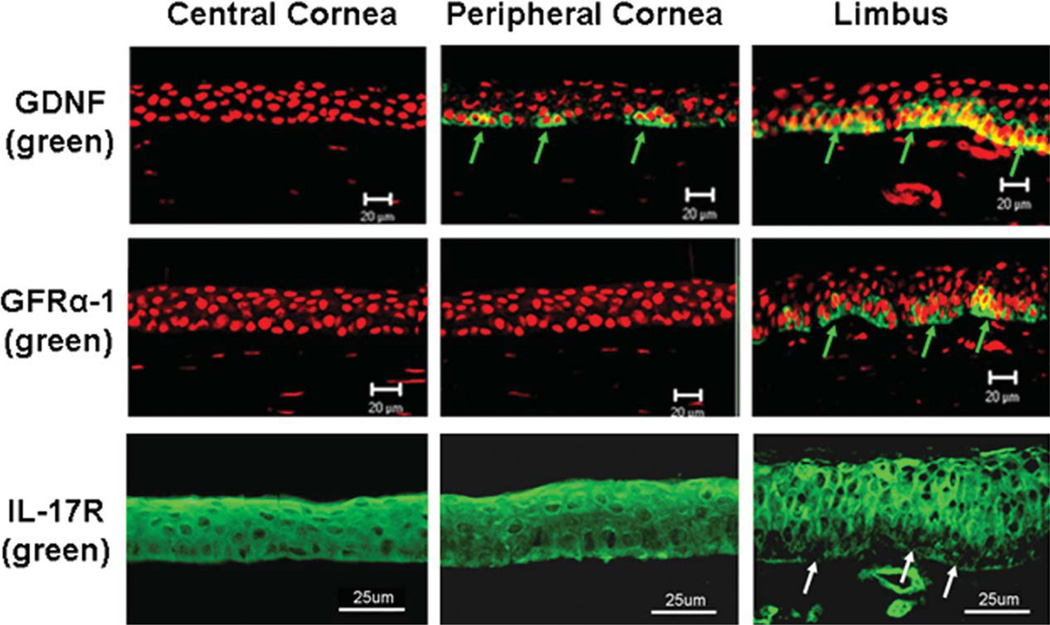
Figure 1.
Representative images showing the immunofluorescent staining of GDNF, GFRα-1, and IL-17R (green color) with propidium iodide (red) nuclear counterstaining on frozen sections of human corneoscleral tissues. Green arrows: positive signals for GDNF or GFRα-1; White arrows: negative staining for IL-17R. Scale bar = 20 or 25 µm. Abbreviations: GDNF, glial cell-derived neurotrophic factor; GFRα-1, GDNF family receptor α-1; IL, interleukin.

