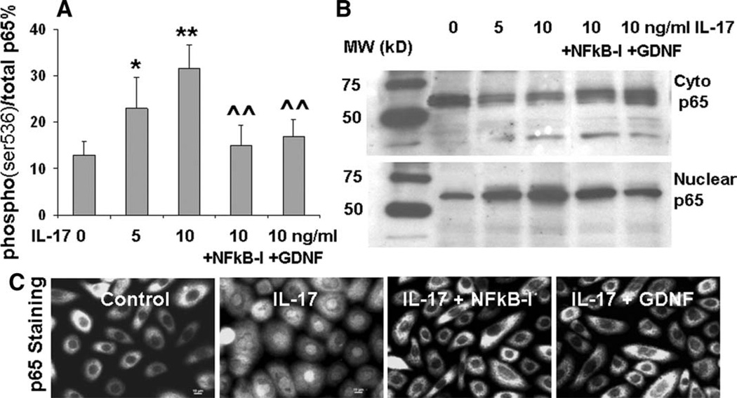
Figure 4.
NF-κB activation in IL-17 stimulated inflammatory responses by human limbal epithelial cells (HLECs). The HLECs were preincubated with 5 µM quinazoline NF-κB-I or 10 ng/ml GDNF for 1 hour before adding 5–10 ng/ml IL-17A. (A): The cells in 96-well plates treated for 30 minutes were used for cell-based enzyme-linked immunosorbent assay quantification of p65 (ser536) phosphorylation (% Phospho-/total p65). Results shown are mean ± SD of three independent experiments, *, p < .05. **, p < .01 compared with controls; ^, p < .05. ^^, p < .01 compared with IL-17 stimulated levels, by analysis of variance (ANOVA) test. (B): The cells in six-well plates treated for 4 hours were subjected to cytoplasm and nuclear protein extraction to evaluate NF-κB p65 nuclear translocation by Western blot analysis. (C): The cells seeded in eight-chamber slides were fixed by methanol for immunofluorescent staining with rabbit anti-human p65 antibody and Alexa-Fluor 488-conjugated second antibodies. The images were representative of results obtained in three independent experiments. Abbreviations: C-p65, cytoplasm p65; GDNF, glial cell-derived neurotrophic factor; IL, interleukin; NF-κB-I, NF-κB inhibitor.

