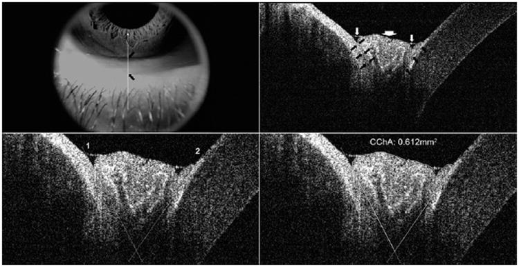
Figure 2.
Measurement of cross-sectional conjunctivochalasis are a (CChA) using RTVue-100. (Top left) Appearance of inferior eyelid and cornea where the vertical scans were taken. Black arrows mark the upper and lower margins of conjunctivochalasis protruding over the lid margin. (Top right) Uniform triangular-shaped tear meniscus between the cornea and lower lid is not visualized in this image; rather, 2 separate small tear menisci on either side of the chalasis are noted (white arrows). The white arrowhead points to the conjunctivochalasis. After the image was digitally magnified 3 times, tissue boundaries (black arrows) among the lower lid, prolapsed conjunctiva, and bulbar conjunctiva/inferior part of the cornea were discriminated from each based on the different levels of the brightness between tissues. (Bottom left) Based on previous determined boundaries, intersecting lines (numbered 1 and 2) were placed to outline the conjunctiva protruding into the junction of the lower lid margin and cornea. (Bottom right) Cross-sectional area of the outlined conjunctivochalasis was measured as 0.612 mm2 using the instrument's software.

