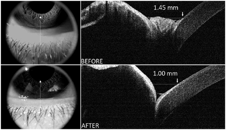
Figure 7.
An example of AS-OCT images of conjunctivochalasis located centrally before (Top right and left) and after (Bottom right and left) thermocauterization. Images show the conjunctivochalasis protruding into lower central tear meniscus shrunk after cauterization and a uniform tear meniscus was restored. The distance between landmarks on the lower cornea and lower lid was 1.45 mm prior to cauterization of severe conjunctivochalasis, decreasing to 1.00 mm after conjunctival cauterization.

