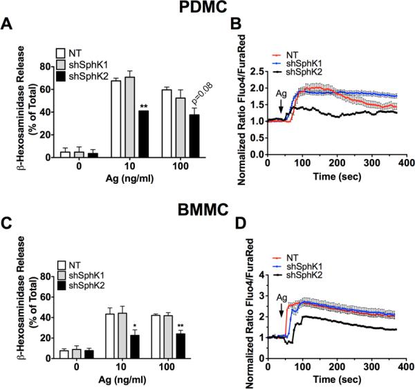
Figure 3. shRNA-mediated silencing of SphK2, but not SphK1, results in impaired degranulation and calcium responses in PDMC and BMMC.

Lentivirus transduced PDMC (A, B) or BMMC (C, D) were sensitized with anti-DNP IgE as described in methods. Cells were washed and then stimulated with the indicated concentrations of antigen. (A, C) Degranulation was calculated as the percentage of β-hexosaminidase released to the incubation media as compared to the total β-hexosaminidase cellular content. Data shown are the average of 3-5 independent cultures done in triplicate ± S.E. Statistical significance compared to nontarget control, * P<0.05, **P<0.01. (B, D) Lentivirus transduced cells were loaded with Fluo4-AM and FuraRed-AM. Following antigen (Ag) stimulation, calcium responses were measured using confocal microscopy and are shown as a ratio of intensity of Fluo4-AM/FuraRed-AM. One representative experiment is shown averaging 15 to 20 individual cells/ time point. Data shown are a mean ± S.E. Similar results were obtained in 3 independent cultures measuring calcium as the ratio of emission at 510 nm when FURA-2AM loaded cells were excited at 340 and 380 nm in populational analysis (data not shown).
