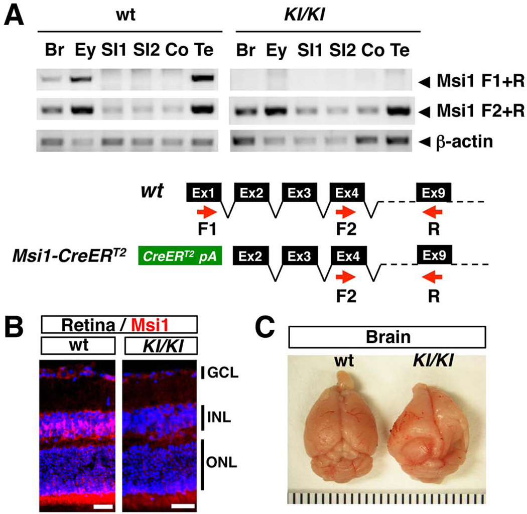
FIG. 2. Expression of Msi1-CreERT2 in homozygous knock-in mice.
(A) RT-PCR analysis of Msi1 mRNA expression in wild type (wt) and Msi1-CreERT2 homozygous knock-in mice (KI/KI). Red arrows, PCR primers; Br, brain; Ey, eye; SI, small intestine; Co, colon; Te, testis. (B) Msi1 expression was detected in Muller glial cells in the INL and photoreceptors in the ONL of the wild type (wt) retina, but not in the retina of homozygous knock-in mice (KI/KI). GCL, ganglion cell layer; INL, inner nuclear layer; ONL, outer nuclear layer. The nucleus is counterstained with DAPI. Bars represent 50 µm. (C) Hydrocephalus was often observed in Msi1-CreERT2 homozygous knock-in mice (KI/KI).

