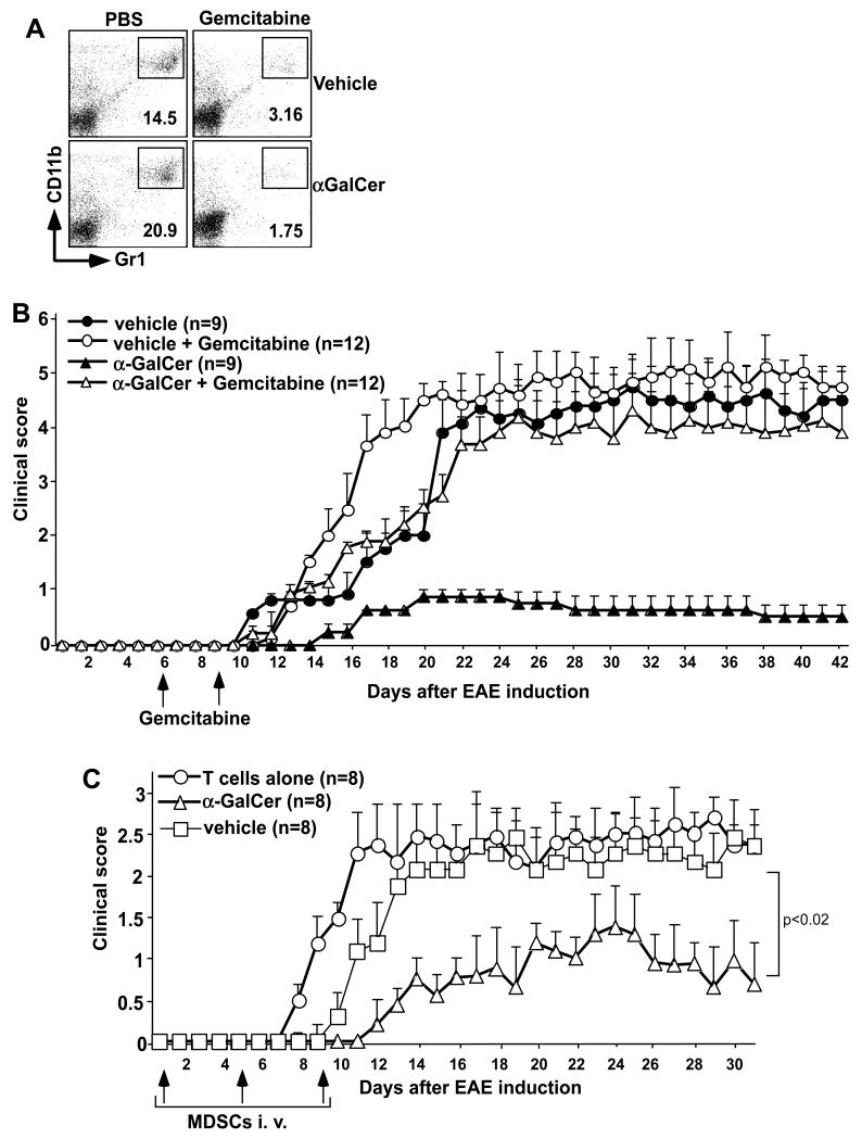
FIGURE 4.
Effects of MDSC depletion or adoptive transfer on EAE. (A,B) EAE was induced in B6 mice and these animals were treated with vehicle or α-GalCer. Mice were then treated at days 6 and 9 after EAE induction with PBS or gemcitabine at 20 mg/kg body weight by i.p. injection. (A) At day 11 after EAE induction, spleen cells were analyzed for the prevalence of MDSCs. Note that >80% depletion was observed. (B) EAE clinical scores were determined as described in Methods. (C) Mice induced with aEAE were treated with vehicle or α-GalCer and sacrificed 11 days after EAE induction. MDSCs were enriched from the spleen using magnetic sorting, pulsed with MOGp at 100 μg/ml for 1 hr and 5×10⁶ cells were adoptively transferred into B6 mice on days 1, 4 and 9 following induction of pEAE with MOGp-specific T cells. EAE clinical scores were determined as described for active EAE. Combined data for two experiments with 4 mice in each group are shown.

