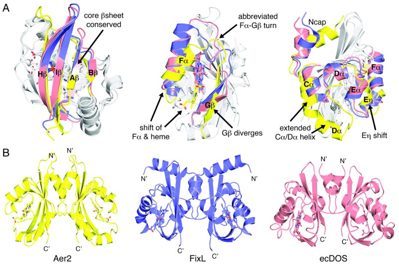
Figure 2. Aer2 is a novel heme-binding PAS domain that forms a parallel PAS dimer.
(A) Structural alignment of the heme-binding PAS domains of Aer2 (yellow, pdb 4HI4), FixL (purple, pdb 1D06), and EcDOS (pink, pdb 1V9Z). Aer2 conserves the core β– sheet (left) but adopts a novel conformation between the Cα helix and Gβ strand elements (middle and right) to bind heme in a unique way. (B) The ferric Aer2 PAS domain forms a parallel dimer with the Ncap and β sheet at the interface. FixL and EcDOS form similar parallel dimers but with different orientations of the PAS domains.

