
Figure 3. Sequence alignment of Aer2 with other heme-binding PAS domains.
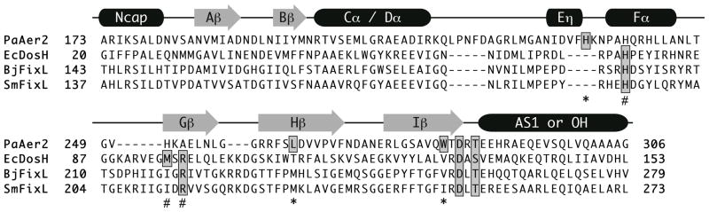
Sequence alignment of PaAer2, EcDos, B. japonicum FixL (BjFixL), and S. meliloti FixL (SmFixL) with secondary structure elements above. The proximal heme-coordinating histidine and distal ligand stabilizing residues are highlighted in grey. Residues important for PaAer2 are denoted by an asterick (*) symbol, while those for EcDOS/FixL are denoted by a # symbol. The C-terminal DxT motif is highlighted in grey.