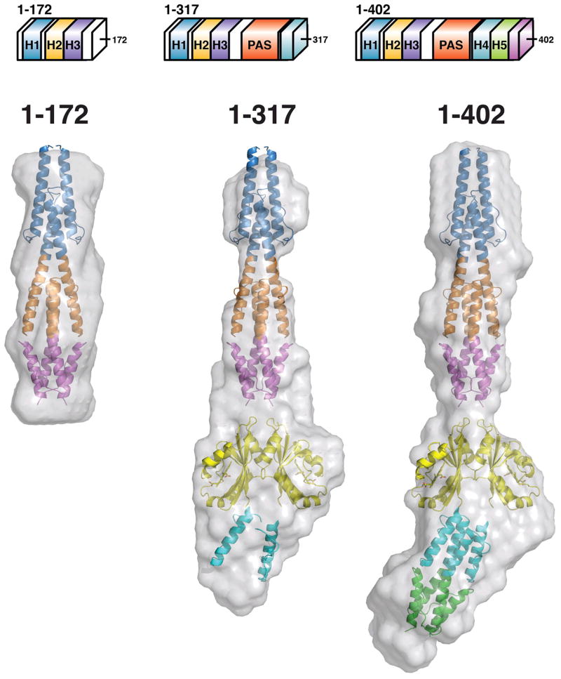
Figure 7. Ab initio SAXS reconstructions of Aer2 protein fragments.
Molecular envelopes (grey) generated from scattering data of Aer2 protein fragments 1–172 (HAMP1–2/3), 1–317 (HAMP1-2/3, PAS, and HAMP4 AS-1/connector), and 1–402 (HAMP1-2/3, PAS, HAMP4/5, and 20 residues of SD). Due to the elongated nature of Aer2, protein fragments of increasing MW were used to allow clear identification of quaternary structure. Crystal structures of Aer2 HAMP1-2/3 (pdb3LNR) and the PAS dimer (pdb 4HI4) were manually placed and fit well inside the envelopes. HAMP2/3 of Aer2 was used as a model for HAMP4/5.

