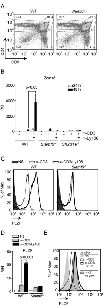
Figure 2. Ly108 is required for full induction of PLZF in vitro.

(A) Characterization of Ly108-deficient (Slamf6−/−) mice. Representative flow plots of thymocyte populations from WT and Slamf6−/− mice stained for CD4 and CD8. Frequencies and absolute numbers of thymocyte populations (N=6) stained for DP, DN CD4+ and CD8+ cells are depicted in Table 1. (B) Zbtb16 expression in WT, Slamf6−/− and SAP-deficient (Sh2d1a−/−) thymocytes stimulated as in Fig.1. Values were normalized to NS WT samples. Similar results were obtained using freshly isolated PS-DP cells as controls (Supplemental Fig. S2A). Data are the means ± SEM of 3 independent experiments, using cells from ≥3 mice each. (C) Representative histograms of intracellular PLZF staining in PS-DP thymocytes from WT and Slamf6−/− mice, NS (solid histogram) or stimulated with plate-bound αCD3±αLy108 for 48 h. Staining controls are shown in Supplemental Fig. S2E. (D) MFI ± SD of PLZF from 3 mice per genotype. (E) Intracellular PLZF expression in PS-DP thymocytes stimulated as in (C) compared to PLZF+CD1d-PBS57 tetramer+ iNKT cells. Histograms are representative of triplicates for each condition. Data in (E) were generated with a different antibody lot than in (C).
