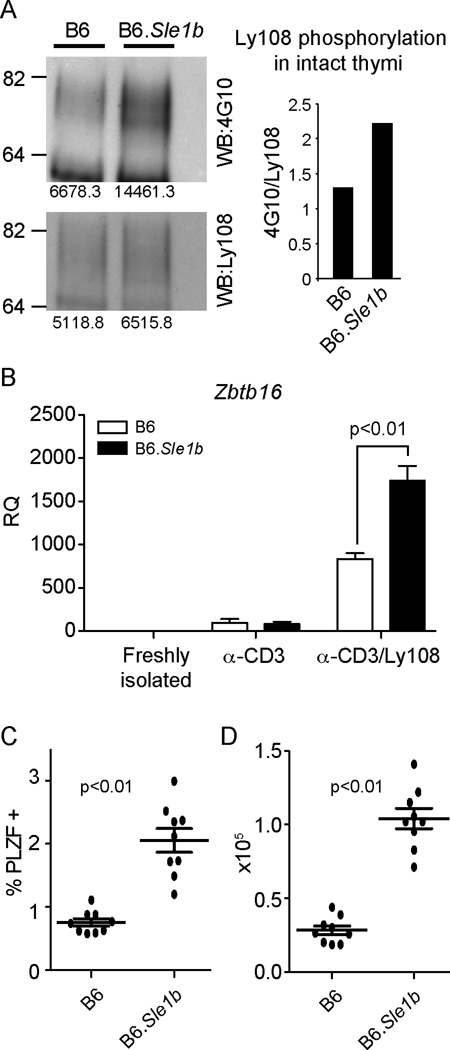
Figure 4. Increased PLZF expressing cells observed in a mouse model with enhanced Ly108 signaling.
(A) Enhanced phosphorylation of Ly108 in intact thymi of B6.Sle1b mice. Ly108 was immunoprecipitated from lysates of intact thymi of C57Bl/6J (B6) and B6.Sle1b mice and probed for phosphotyrosine using 4G10 antibody (top) and for total Ly108 protein (bottom). Quantitation of relative tyrosine phosphorylation is shown on the right. (B) Zbtb16 expression in C57Bl/6J (B6) and B6.Sle1b PS-DP thymocytes stimulated for 48 hrs with plate-bound αCD3±αLy108 as in Figure 1. Data are representative of three independent experiments. Percentages (C) and absolute numbers (D) of PLZF+ CD4+ thymocytes in C57Bl/6J (B6) and B6.Sle1b mice.

