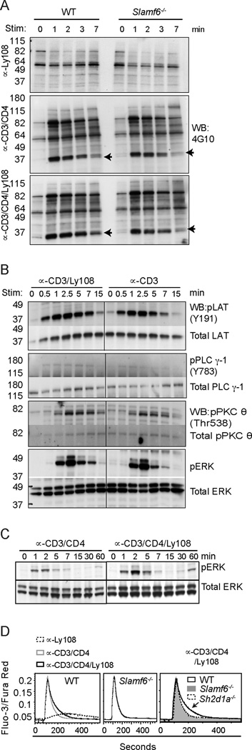
Figure 5. Ly108 costimulation prolongs TCR signaling.

(A) Rested WT and Slamf6−/− thymocytes, pre-incubated with biotinylated αLy108 (top), αCD3+αCD4 (middle), and αCD3+αCD4+αLy108 (bottom), were stimulated with streptavidin for indicated times and lysates immunoblotted for α-phosphotyrosine (4G10). Arrows indicate predicted migration of LAT (left). Quantitation of 37 kDa band corresponding to LAT in (A) is shown in supplemental Fig. S3A (B) WT thymocytes were stimulated with αCD3±αLy108 as in (A) and lysates probed for pLAT (Y191), LAT; pPLCγ-1 (Y783), PLCγ-1; pPKCθ (Thr 538), PKCθ; pERK and ERK. Quantitation of band intensities are shown in supplemental Fig. S3B. (C) WT thymocytes were stimulated with αCD3+αCD4±αLy108 for extended times and probed for phospho and total Erk proteins. (D) Ca2+ flux of WT and Slamf6−/− PS-DP thymocytes stimulated with biotin-conjugated αCD3/CD4±αLy108 followed by streptavidin, as indicated by the ratio of Fluo-3/Fura-Red. Arrow indicates enhanced intracellular Ca2+ levels seen in WT, but not Ly108- or SAP-deficient thymocytes, upon αCD3/CD4/Ly108 stimulation. A–D are representative of 3 independent experiments.
