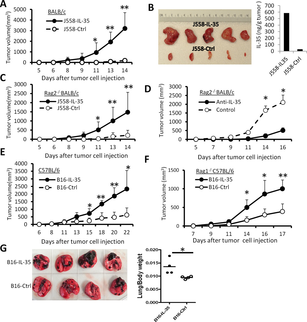
Fig.3. Expression of IL-35 in the tumor microenvironment enhances tumorigenesis.
5 × 10⁶ J558-IL-35 or J558-Ctrl cells were injected into each BALB/c (A) or Rag2−/−BALB/c mouse (C) s.c. The tumor growth was observed over time, and at the end of experiments, tumors were removed from sacrificed mice and photographed. Shown in photo (B) are J558-IL-35 and J558-Ctrl tumors removed from BALB/c mice. ELISA was used to quantify IL-35 concentration in lysates of representative tumors (B). Each Rag2−/−BALB/c mouse was inoculated with 5 × 10⁶ J558-IL-35 cells s.c. in the presence of anti-IL-35 (V1.4C4.22, Shenandoah Biotechnology) or an isotype-matched control mAb (IgG2b, BioXcell) at a concentration of 50 µg/ml. Mice were observed for tumor growth over time. Bars indicate SD of 3 mice in each group and data shown represent two experiments with similar results. 1 × 10⁵ B16-IL-35 or B16-Ctrl cells were injected into each C57BL/6 (E) or Rag1−/−C57BL/6 mouse (F) s.c. The tumor growth was observed over time. Bars in A, C, D, E and F indicate SD of 5 mice in each group. Data shown represents three to five experiments with similar results. (G) 1 × 10⁵ B16-IL-35 or B16-Ctrl cells were injected into each C57BL/6 i.v. Eighteen days after tumor cell injection, lungs from the recipient mice were removed, photographed (left) and weighed, and lung/body weight ratios were calculated and plotted in the right panel. *P<0.05; **P<0.01 by student’s t test.

