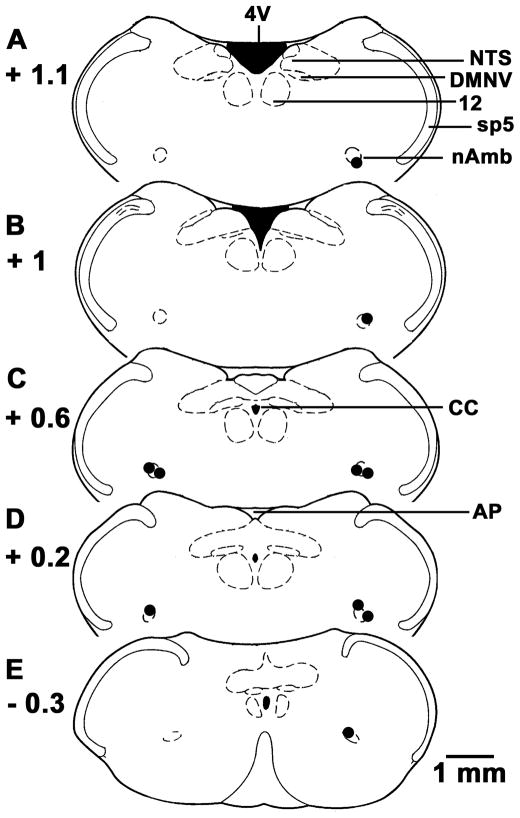
Fig. 6. Histological identification of microinjection sites.
A–E, Drawings of coronal sections at levels 1.1 mm rostral to 0.3 mm caudal to the calamus scriptorius (CS). Microinjection sites are shown as dark spots; each spot represents a site in one animal (n = 10). Abbreviations: AP, area postrema; CC, central canal; DMNV, dorsal motor nucleus of vagus; nAmb, nucleus ambiguus; NTS, nucleus tractus solitarius; Sp5, spinal trigeminal tract; 4V, fourth ventricle; 12, hypoglossal nucleus.

