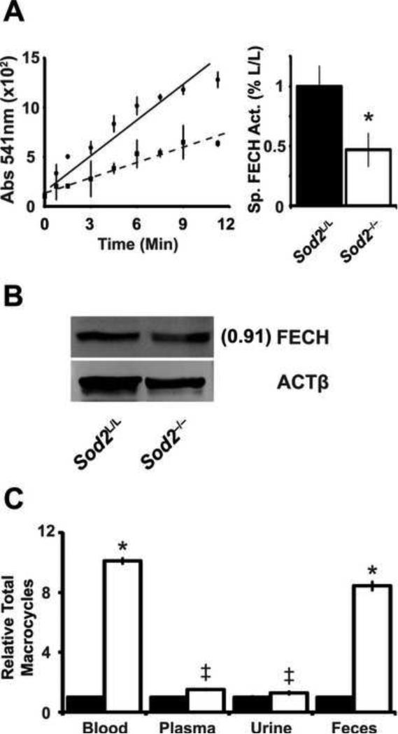
Figure. 5. Ferrochelatase activity is decreased by increased mitochondrial superoxide.
(A) Ferrochelatase (FECH) activity assay performed on bone marrow extracted from 6-week old animals. Left, data displayed as absorbance of zinc-incorporated porphyrin over time. Solid line indicates Sod2L/L, dotted line indicates Sod2-/-. Right, quantification of FECH activity. Specific activity calculated based on relative FECH protein levels (panel B). Three mice of respective genotypes were analyzed per experiment; data are shown as mean and s.d. Where applicable, * = p<0.01 or ‡ = p<0.05 by Student's t-test versus Sod2L/L. (B) Western blot analysis for ferrochelatase (FECH) protein in bone marrow from 6-week old Sod2L/L or Sod2-/- mice. β-actin (ACTβ) shown as loading control. Quantification normalized to ACTβ, then to Sod2L/L using ImageJ software. (C) Spectrophotometric assay of iron-free macrocycles in various tissues in 6-week old Sod2L/L or Sod2-/- mice. Closed bars indicate Sod2L/L, open bars indicate Sod2-/-. Data normalized to respective Sod2L/L tissue.

