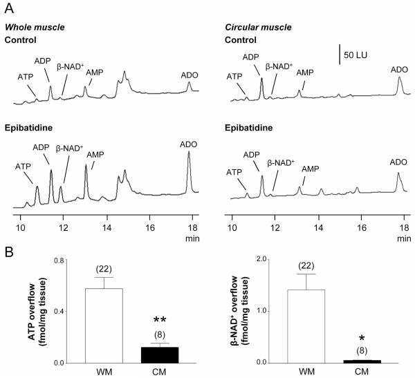
Fig. 2. Release of ATP and β-NAD+ during nAChR stimulation in whole muscle (WM) and circular muscle (CM) preparations of monkey colon.
(A) Chromatograms of tissue superfusates collected before (control) and during nAChR stimulation with epibatidine (500 μM, 30 s) in WM and CM preparations of monkey colon. Small amounts of ATP and β-NAD+ were present in WM and CM superfusates in the absence of agonist. Epibatidine evoked release of ATP and β-NAD+ in WM preparations, but not in CM preparations. Scale applies to all chromatograms. LU, luminescence units. (B) Averaged data are means ± SEM, showing epibatidine-evoked release of ATP (left) and β-NAD+ (right) from monkey WM and CM preparations. Overflow (femtomoles per milligram of tissue) is the overflow during nAChR activation less spontaneous overflow. Overflow of ATP and β-NAD+ was significantly less in CM preparations. Asterisks denote significant differences from WM release (*P < 0.05, **P < 0.01); number of experiments in parenthesis.

