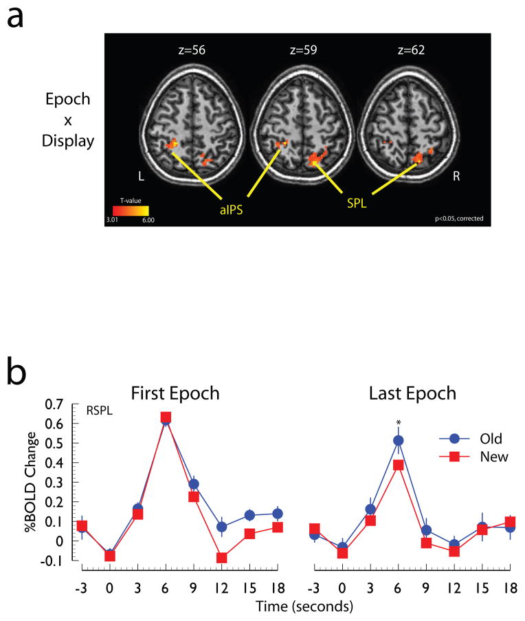
Figure 3. Evidence for modulation of parietal cortex responses.
Panel A: Regions of parietal cortex sensitive to the Epoch x Display interaction. Panel B: Time course of the hemodynamic response of the SPL local maximum shown in panel a. Abbreviations: SPL, superior parietal lobe; IPS, intraparietal sulcus, IPS; a, anterior; R, right; L, left.

