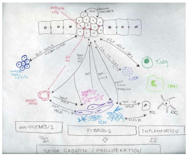
Figure 3.
Anatomical and cellular alterations leading to HCC development. (a) Normal liver parenchyma. Hepatocytes with microvilli and fenestrated sinusoidal cells that favours the metabolic exchange. Space of Disse with few quiescence stellate cells containing lipid droplets. (b) Fibrotic liver. Upon chronic liver injury, hepatocytes loose the microvilli and sinusoids their fenestration, stellate cells become activated, loosing the lipid droplets and secreting ECM. (c) Hepaocellular carcinoma. Malignant transformation of hepatocytes with uncontrolled growth. Infiltration of inflammatory cells and cytokines. Development of new vessels (neoangiogenesis) and extense fibrosis with recruitment of tumor associated fibroblasts and cancer stem cells.

